CORRELATED DIFFUSION IMAGING
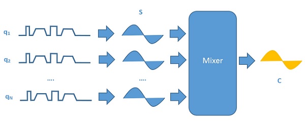
We introduce a new form of diffusion magnetic resonance imaging called correlated diffusion imaging, where the tissue being imaged is characterized by the joint correlation of diffusion signal attenuation across multiple gradient pulse strengths and timings.
To characterize the tissue being imaged, the acquired signals are mixed together to obtain the local correlation of signal attenuation across the acquired signals. By taking into account signal attenuation at different water diffusion motion sensitivities, correlated diffusion imaging can provide improved delineation between cancerous tissue and healthy tissue when compared to existing diffusion imaging modalities.
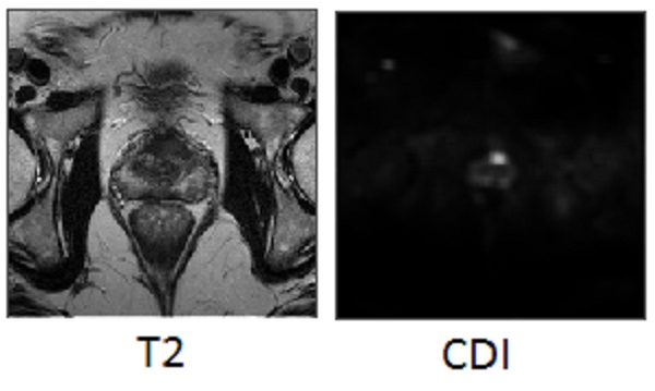
An example of the use of correlated diffusion imaging for prostate imaging can be seen below. It can be seen that the tumor stands out well (see bright spot) on correlated diffusion imaging (right) and not at all on T2-weighted imaging (left) in a patient with prostate cancer. This illustrates the potential of correlated diffusion imaging as a powerful tool for cancer diagnosis and treatment.
For more information, please read:
1. A. Wong, J. Glaister, A. Cameron, and M. Haider, Correlated Diffusion Imaging. BMC Med Imaging. 2013 Aug 8;13:26. doi: 10.1186/1471-2342-13-26.
2. F. Khalvati, J. Zhang, M. Haider, and A. Wong, Enhanced Dual-Stage Correlated Diffusion Imaging. Annual International Conference of the IEEE Engineering in Medicine and Biology Society (IEEE EMBC). 2016.
3. A. Wong, F. Khalvati, and M. Haider, Dual-stage correlated diffusion imaging. IEEE ISBI. 2015 Apr 16-19; 75-78.
4. J. Zhang, A. Eilaghi, M. Haider, A. Wong, and F. Khalvati, Optimized Correlated Diffusion Imaging for Prostate Cancer Detection. Annual Meeting of the Imaging Network of Ontario. 2016.
5. A. Wong, Correlated Diffusion Imaging System and Method for Identification of Biological Tissue of Interest. United States Patent Application 14/309,318.
6. A. Wong, Correlated Diffusion Imaging System and Method for Identification of Biological Tissue of Interest. Canadian Patent Application 2,854,844.
We introduce a new form of diffusion magnetic resonance imaging called correlated diffusion imaging, where the tissue being imaged is characterized by the joint correlation of diffusion signal attenuation across multiple gradient pulse strengths and timings.
To characterize the tissue being imaged, the acquired signals are mixed together to obtain the local correlation of signal attenuation across the acquired signals. By taking into account signal attenuation at different water diffusion motion sensitivities, correlated diffusion imaging can provide improved delineation between cancerous tissue and healthy tissue when compared to existing diffusion imaging modalities.
An example of the use of correlated diffusion imaging for prostate imaging can be seen below. It can be seen that the tumor stands out well (see bright spot) on correlated diffusion imaging (right) and not at all on T2-weighted imaging (left) in a patient with prostate cancer. This illustrates the potential of correlated diffusion imaging as a powerful tool for cancer diagnosis and treatment.
For more information, please read:
1. A. Wong, J. Glaister, A. Cameron, and M. Haider, Correlated Diffusion Imaging. BMC Med Imaging. 2013 Aug 8;13:26. doi: 10.1186/1471-2342-13-26.
2. F. Khalvati, J. Zhang, M. Haider, and A. Wong, Enhanced Dual-Stage Correlated Diffusion Imaging. Annual International Conference of the IEEE Engineering in Medicine and Biology Society (IEEE EMBC). 2016.
3. A. Wong, F. Khalvati, and M. Haider, Dual-stage correlated diffusion imaging. IEEE ISBI. 2015 Apr 16-19; 75-78.
4. J. Zhang, A. Eilaghi, M. Haider, A. Wong, and F. Khalvati, Optimized Correlated Diffusion Imaging for Prostate Cancer Detection. Annual Meeting of the Imaging Network of Ontario. 2016.
5. A. Wong, Correlated Diffusion Imaging System and Method for Identification of Biological Tissue of Interest. United States Patent Application 14/309,318.
6. A. Wong, Correlated Diffusion Imaging System and Method for Identification of Biological Tissue of Interest. Canadian Patent Application 2,854,844.