COMPENSATED MAGNETIC RESONANCE IMAGING
We introduce a novel compensated magnetic resonance imaging (cMRI) for significantly improving tissue detail and contrast for the purpose of clinical screening and diagnosis. The novel compensated magnetic resonance imaging system incorporates the intrinsic properties of the MRI apparatus to compensate for artifacts and degradation caused as a result of the imaging process.
By taking into account the different properties and characteristics of the MRI imaging system and process, compensated magnetic resonance imaging can produce MRI imagery with significantly improved tissue detail and contrast compared to existing MRI systems, which is very useful for providing more information for diagnostic purposes.
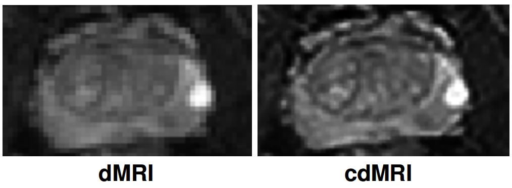
An example of the use of compensated magnetic resonance imaging for prostate imaging can be seen below. It can be seen that the tissue detail and contrast is signficantly improved in the prostate gland of the diffusion MRI image achieved using compensated magnetic resonance imaging (right) when compared to that produced using a standard MRI system (left). This illustrates the potential of compensated magnetic resonance imaging as a powerful tool for cancer diagnosis and treatment.
For more information, please read:
1. A. Boroomand, E. Li, M. Shafiee, F. Khalvati, M. Haider, and A. Wong, Bayesian-based Compensated Magnetic Resonance Imaging. Annual International Conference of the IEEE Engineering in Medicine and Biology Society (IEEE EMBC). 2016.
2. A. Boroomand, E. Li, M. Shafiee, F. Khalvati, M. Haider, and A. Wong, A Unified Reconstruction Framework for Compensated Magnetic Resonance Imaging. Annual Meeting of the Imaging Network of Ontario. 2016.
3. E. Li, M. Shafiee, A. Boroomand, F. Khalvati, M. Haider, and A. Wong, Compensated Diffusion Magnetic Resonance Imaging. Vision Letters. 2015.
4. A. Wong, A compensated magnetic resonance imaging system and method for improved magnetic resonance imaging and diffusion imaging quality. United States Patent Application 14/309,318.
We introduce a novel compensated magnetic resonance imaging (cMRI) for significantly improving tissue detail and contrast for the purpose of clinical screening and diagnosis. The novel compensated magnetic resonance imaging system incorporates the intrinsic properties of the MRI apparatus to compensate for artifacts and degradation caused as a result of the imaging process.
By taking into account the different properties and characteristics of the MRI imaging system and process, compensated magnetic resonance imaging can produce MRI imagery with significantly improved tissue detail and contrast compared to existing MRI systems, which is very useful for providing more information for diagnostic purposes.
An example of the use of compensated magnetic resonance imaging for prostate imaging can be seen below. It can be seen that the tissue detail and contrast is signficantly improved in the prostate gland of the diffusion MRI image achieved using compensated magnetic resonance imaging (right) when compared to that produced using a standard MRI system (left). This illustrates the potential of compensated magnetic resonance imaging as a powerful tool for cancer diagnosis and treatment.
For more information, please read:
1. A. Boroomand, E. Li, M. Shafiee, F. Khalvati, M. Haider, and A. Wong, Bayesian-based Compensated Magnetic Resonance Imaging. Annual International Conference of the IEEE Engineering in Medicine and Biology Society (IEEE EMBC). 2016.
2. A. Boroomand, E. Li, M. Shafiee, F. Khalvati, M. Haider, and A. Wong, A Unified Reconstruction Framework for Compensated Magnetic Resonance Imaging. Annual Meeting of the Imaging Network of Ontario. 2016.
3. E. Li, M. Shafiee, A. Boroomand, F. Khalvati, M. Haider, and A. Wong, Compensated Diffusion Magnetic Resonance Imaging. Vision Letters. 2015.
4. A. Wong, A compensated magnetic resonance imaging system and method for improved magnetic resonance imaging and diffusion imaging quality. United States Patent Application 14/309,318.