COMPENSATED ULTRASOUND IMAGING
We introduce a novel compensated ultrasound imaging (cUI) for significantly improving contrast, signal-to-noise-ratio, and significantly reduced aberrations for the purpose of clinical screening and diagnosis. The novel compensated ultrasound imaging system incorporates the intrinsic properties of the ultrasound apparatus to compensate for artifacts and degradation caused as a result of the imaging process.
By taking into account the different properties and characteristics of the ultrasound imaging system and process, compensated ultrasound imaging can produce US imagery with significantly improved detail, contrast, and signficantly reduced aberrations compared to existing ultrasound systems, which is very useful for providing more accurate information for diagnostic purposes.
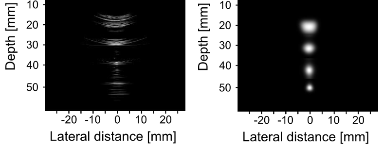
An example of the use of compensated ultrasound imaging on a ultrasound target can be seen below. It can be seen that the detail, contrast, and the shape of the dots in the image is signficantly improved using compensated ultrasound imaging (right) when compared to that produced using a standard ultrasound imaging system (left). This illustrates the potential of compensated ultrasound imaging as a powerful tool for diagnosis and therapy guidance.
For more information, please read:
1. I. Ben Daya, A. Chen, J. Yeow, and A. Wong, Compensated Row-Column Ultrasound Imaging System Using Fisher Tippett Multilayered Conditional Random Field Model. PLoS ONE. 2015.
We introduce a novel compensated ultrasound imaging (cUI) for significantly improving contrast, signal-to-noise-ratio, and significantly reduced aberrations for the purpose of clinical screening and diagnosis. The novel compensated ultrasound imaging system incorporates the intrinsic properties of the ultrasound apparatus to compensate for artifacts and degradation caused as a result of the imaging process.
By taking into account the different properties and characteristics of the ultrasound imaging system and process, compensated ultrasound imaging can produce US imagery with significantly improved detail, contrast, and signficantly reduced aberrations compared to existing ultrasound systems, which is very useful for providing more accurate information for diagnostic purposes.
An example of the use of compensated ultrasound imaging on a ultrasound target can be seen below. It can be seen that the detail, contrast, and the shape of the dots in the image is signficantly improved using compensated ultrasound imaging (right) when compared to that produced using a standard ultrasound imaging system (left). This illustrates the potential of compensated ultrasound imaging as a powerful tool for diagnosis and therapy guidance.
For more information, please read:
1. I. Ben Daya, A. Chen, J. Yeow, and A. Wong, Compensated Row-Column Ultrasound Imaging System Using Fisher Tippett Multilayered Conditional Random Field Model. PLoS ONE. 2015.