SPECTRAL LIGHT-FIELD FUSION MICRO-TOMOGRAPHY
We introduce a new form of light-field imaging called spectral light-field fusion micro-tomography (SLFM), where an innovative, highly-compact lensfree pulsed lighting and detection apparatus captures interferometric light-field encodings at different wavelengths, and a Bayesian-based fusion strategy facilitates for the reconstruction of a high-resolution, high-contrasted fused complex object light-field from the encodings. It is from this fused complex object light-field that we obtain tomograms of the imaged specimen. By fusing unique object light-field information captured at different wavelengths, SLFM can achieve 4D label-free tomographic imaging of biological specimens in a very low-cost, compact form factor without using any lenses, while achieving nanometer resolutions with a ultra-wide field-of-view (>100 times greater than 40X optical microscopy systems) and quantitative phase contrast information.
For more information on the earlier variant of SLFM (we have since achieved micro-tomography at greater than five times the effective resolution), please read:
1. F. Kazemzadeh and A. Wong, Laser light-field fusion for wide-field lensfree on-chip phase contrast nanoscopy. Nature Scientific Reports, 2016.
2. F. Kazemzadeh, E. Kuang, and A. Wong, Compact, Field-Portable Lens-free Microscope using Superresolution Spatio-Spectral Light-field Fusion. Journal of Comptuational Vision and Imaging Systems, 2016.
3. F. Kazemzadeh, C. Jin, S. Molladavoodi, Y. Mei, M. Emelko, M. Gorbet, and A. Wong, Lensfree Spectral Light-field Fusion Microscopy for Contrast- and Resolution-enhanced Imaging of Biological Specimens. Optics Letters, 2015.
4. F. Kazemzadeh and A. Wong, Lens-free Multi-Laser Spectral Light-Field Fusion Microscopy. Journal of Computational Vision and Imaging Systems, 2015.
5. A. Wong, F. Kazemzadeh, C. Jin, and X. Wang, Lensfree Bayesian-based aberration correction and numerical diffraction for improved lensfree on-chip microscopy of biological specimens. Optics Letters, 2015.
6. F. Kazemzadeh and A. Wong, Whole-Slide Digital Pathology via Lens-free Spectral Light-field Fusion Microscopy. Annual Meeting of the Imaging Network of Ontario. 2016.
7. F. Kazemzadeh and A. Wong, A System, Method, and Apparatus for Ultra-resolved Ultra-wide Field-of-view Multispectral and Hyperspectral Holographic Microscopy. United States Patent Application 62/155,416.
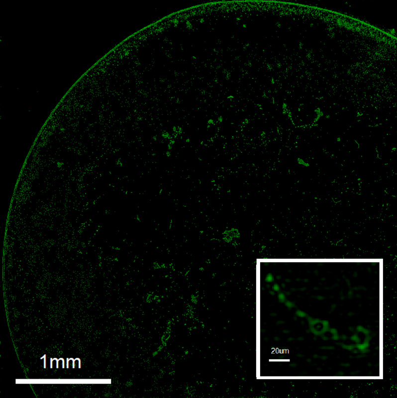
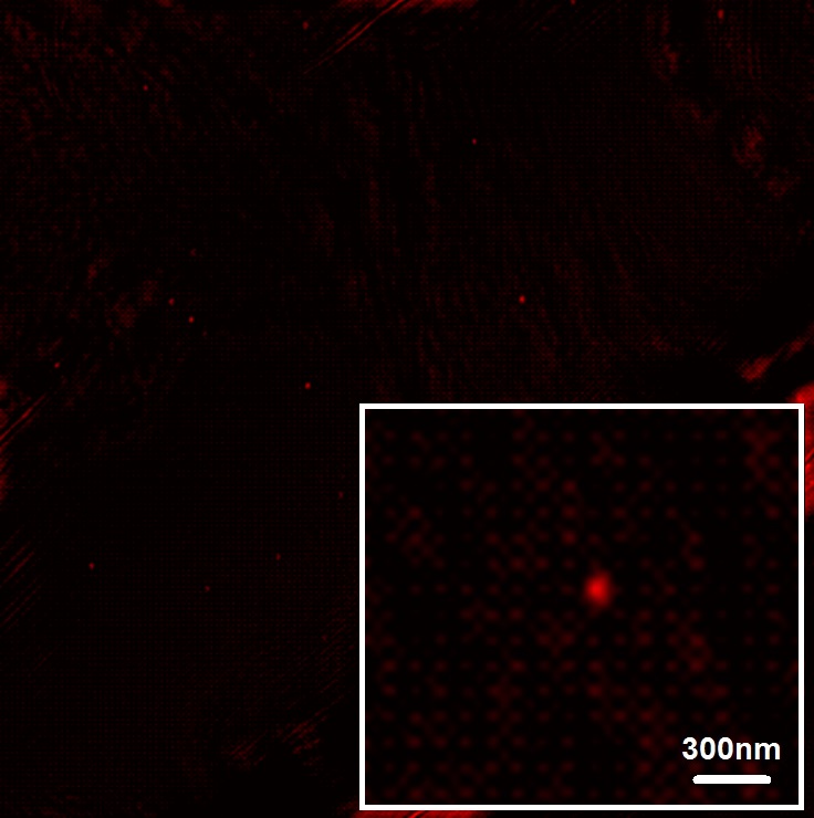
Example images captured of using our current SLFM system (top-left: phase contrast ROI of sonicated Anabaena flos-aquae, top-right: phase contrast ROI of silica microspheres, middle: intensity projection of lung tissue, bottom: intensity projection of bee head):


We introduce a new form of light-field imaging called spectral light-field fusion micro-tomography (SLFM), where an innovative, highly-compact lensfree pulsed lighting and detection apparatus captures interferometric light-field encodings at different wavelengths, and a Bayesian-based fusion strategy facilitates for the reconstruction of a high-resolution, high-contrasted fused complex object light-field from the encodings. It is from this fused complex object light-field that we obtain tomograms of the imaged specimen. By fusing unique object light-field information captured at different wavelengths, SLFM can achieve 4D label-free tomographic imaging of biological specimens in a very low-cost, compact form factor without using any lenses, while achieving nanometer resolutions with a ultra-wide field-of-view (>100 times greater than 40X optical microscopy systems) and quantitative phase contrast information.
For more information on the earlier variant of SLFM (we have since achieved micro-tomography at greater than five times the effective resolution), please read:
1. F. Kazemzadeh and A. Wong, Laser light-field fusion for wide-field lensfree on-chip phase contrast nanoscopy. Nature Scientific Reports, 2016.
2. F. Kazemzadeh, E. Kuang, and A. Wong, Compact, Field-Portable Lens-free Microscope using Superresolution Spatio-Spectral Light-field Fusion. Journal of Comptuational Vision and Imaging Systems, 2016.
3. F. Kazemzadeh, C. Jin, S. Molladavoodi, Y. Mei, M. Emelko, M. Gorbet, and A. Wong, Lensfree Spectral Light-field Fusion Microscopy for Contrast- and Resolution-enhanced Imaging of Biological Specimens. Optics Letters, 2015.
4. F. Kazemzadeh and A. Wong, Lens-free Multi-Laser Spectral Light-Field Fusion Microscopy. Journal of Computational Vision and Imaging Systems, 2015.
5. A. Wong, F. Kazemzadeh, C. Jin, and X. Wang, Lensfree Bayesian-based aberration correction and numerical diffraction for improved lensfree on-chip microscopy of biological specimens. Optics Letters, 2015.
6. F. Kazemzadeh and A. Wong, Whole-Slide Digital Pathology via Lens-free Spectral Light-field Fusion Microscopy. Annual Meeting of the Imaging Network of Ontario. 2016.
7. F. Kazemzadeh and A. Wong, A System, Method, and Apparatus for Ultra-resolved Ultra-wide Field-of-view Multispectral and Hyperspectral Holographic Microscopy. United States Patent Application 62/155,416.
Example images captured of using our current SLFM system (top-left: phase contrast ROI of sonicated Anabaena flos-aquae, top-right: phase contrast ROI of silica microspheres, middle: intensity projection of lung tissue, bottom: intensity projection of bee head):